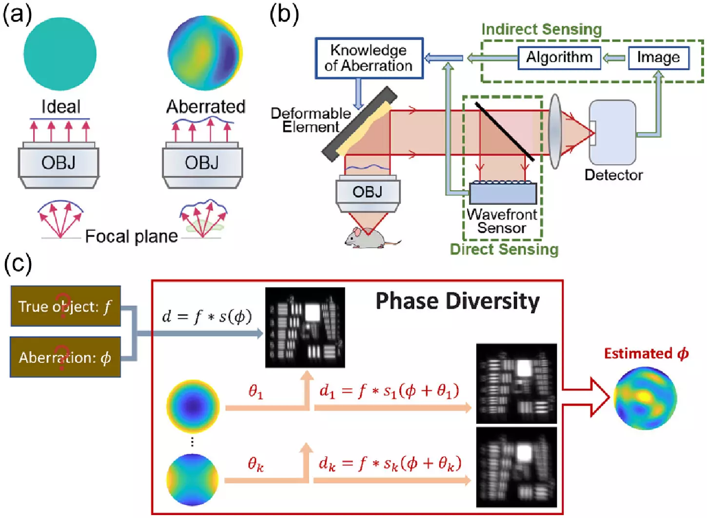
In a groundbreaking study published in the journal Optica, a team of researchers at HHMI’s Janelia Research Campus has introduced a new method to unblur microscopy images, inspired by techniques used in astronomy. This innovative approach promises to provide biologists with faster and more cost-effective solutions for obtaining clearer and sharper images of biological samples.
Microscopists have long grappled with the challenge of image distortion caused by the bending of light in thick biological samples. Similar to how astronomers correct for aberrations in images of far-away galaxies, biologists have explored adaptive optics techniques to enhance the quality of their microscopy images. However, traditional adaptive optics methods have been plagued by complexity, high costs, and slow processing speeds, limiting their accessibility to many research labs.
In their quest to democratize adaptive optics for the life sciences, the researchers at Janelia Research Campus have turned to a set of techniques known as phase diversity. These methods, widely employed in astronomy but relatively new to biology, involve adding images with known aberrations to a blurry image with unknown aberrations. The additional data provided by these images allows for the unblurring of the original image, without requiring significant modifications to the imaging system.
To adapt phase diversity techniques for microscopy, the team first reconfigured an astronomy algorithm and conducted rigorous simulations to validate the approach. They then constructed a microscope equipped with a deformable mirror and additional lenses to introduce known aberrations. These minor modifications, along with enhancements to the software for phase diversity correction, enabled the team to demonstrate a 100-fold increase in the speed of calibrating the deformable mirror compared to existing methods.
In testing the new method, the researchers successfully corrected randomly generated aberrations, resulting in clearer images of fluorescent beads and fixed cells. The next phase of their research involves applying the technique to real-world biological samples, including living cells and tissues, and expanding its utility to more complex microscopy setups. Additionally, the team aims to streamline the method, making it more automated and user-friendly to broaden its accessibility to a wider range of laboratories.
The innovative approach developed by the researchers at Janelia Research Campus offers a promising solution to the longstanding challenge of image distortion in microscopy. By leveraging techniques from astronomy and introducing phase diversity methods to the life sciences, this new method enables biologists to obtain clearer and sharper images of biological samples in a faster and more cost-effective manner. With further refinement and validation, this revolutionary approach has the potential to transform the field of microscopy and empower researchers to gain deeper insights into the intricate world of living organisms.


Leave a Reply