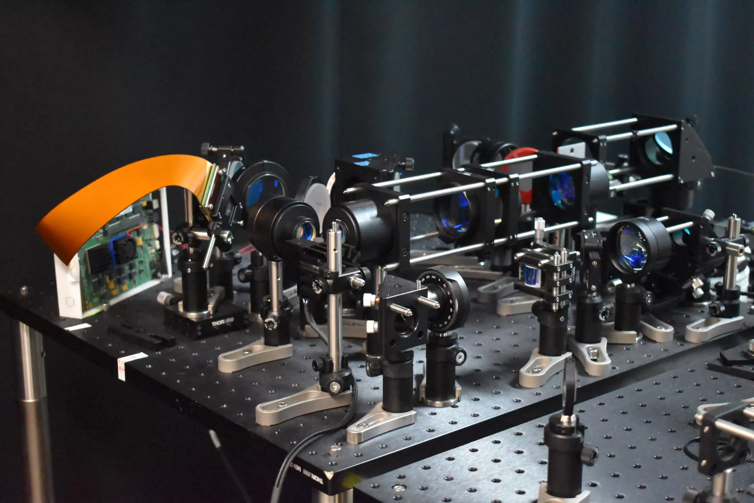
Scientists have recently introduced a groundbreaking two-photon fluorescence microscope that has the capability to capture high-speed images of neural activity at the cellular level. This new technology provides a more efficient and less invasive alternative to traditional two-photon microscopy, offering researchers a clearer understanding of how neurons communicate in real time. By shedding light on these intricate processes, this innovation has the potential to unlock new insights into brain function and neurological disorders.
The newly developed two-photon fluorescence microscope incorporates an innovative adaptive sampling scheme that replaces the conventional point illumination with line illumination. This allows for in vivo imaging of neuronal activity in a mouse cortex at speeds that are ten times faster than those achieved by traditional two-photon microscopy. Moreover, the reduced laser power used on the brain tissue limits potential damage by over tenfold. This groundbreaking method opens up a wealth of possibilities for studying the dynamics of neural networks in real-time, offering invaluable contributions to the fields of learning, memory, and decision-making.
Researchers behind this revolutionary microscope suggest that their technology could hold the key to uncovering pathology at the earliest stages of neurological diseases. By observing neural activity in real-time, this advanced tool offers a unique opportunity to explore the intricacies of conditions such as Alzheimer’s, Parkinson’s, and epilepsy. The ability to study brain function with unprecedented detail and precision could lead to more effective treatments and interventions for these debilitating disorders.
Traditional two-photon microscopy, though capable of providing detailed images, is limited by its slow imaging speed and potential harm to brain tissue. The new adaptive sampling strategy introduced by researchers addresses these constraints by utilizing a line of light to illuminate specific brain regions. By excelling only the neurons of interest rather than inactive areas, the total light energy delivered to the brain tissue is significantly reduced, minimizing the risk of damage. This innovative approach, powered by a digital micromirror device, allows for dynamic shaping and targeting of active neurons, enabling precise and swift imaging of neural processes.
The advancements in the field of microscopy presented in this research pave the way for the study of dynamic neural processes in real time. By combining adaptive line-excitation techniques with cutting-edge computational algorithms, researchers can isolate and monitor individual neurons with unprecedented speed and accuracy. This ability to capture rapid neuronal events at a rate of 198 Hz surpasses the capabilities of traditional microscopes, offering a glimpse into the intricate functions of the brain that would otherwise go unnoticed.
Looking ahead, scientists involved in this project aim to enhance the capabilities of the microscope by integrating voltage imaging features to provide rapid insights into neural activity. They also plan to apply this technology to real neuroscience scenarios, such as monitoring brain activity during learning processes and studying neurological disorders. Furthermore, efforts are underway to improve the usability of the microscope and reduce its size to broaden its applications in neuroscience research. The potential for this revolutionary two-photon fluorescence microscope to revolutionize our understanding of the brain and its functions cannot be understated.


Leave a Reply